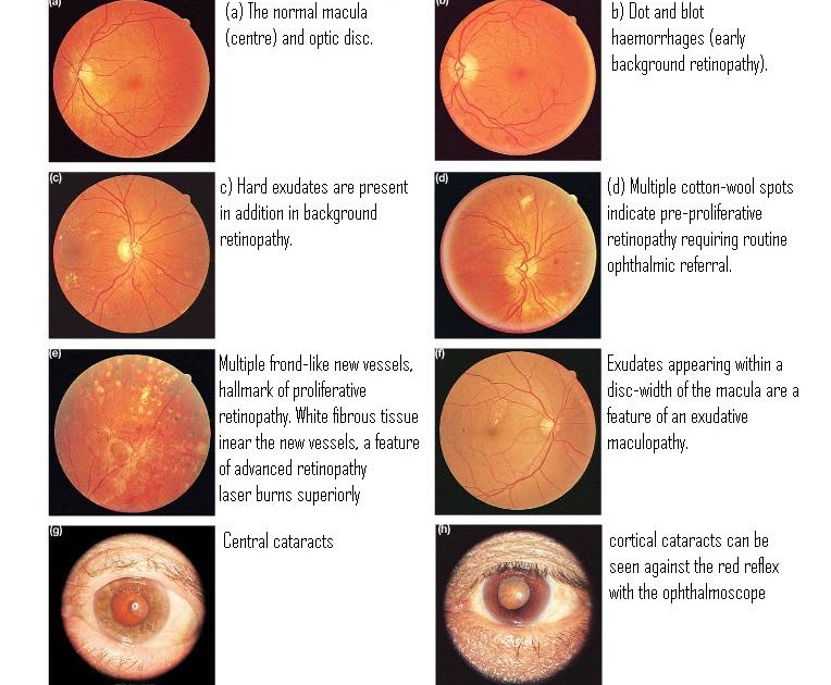
More severe vision loss can occur if the macula or large areas of the retina are detached. Macular wrinkling can cause visual distortion. Traction retinal detachment: When PDR is present, scar tissue associated with neovascularization can shrink, wrinkling and pulling the retina from its normal position. When the blood clears, vision may return to its former level unless the macula is damaged. Vitreous haemorrhage alone does not cause permanent vision loss. If the eye does not clear the vitreous blood adequately within a reasonable time, vitrectomy surgery may be recommended. It may take days, months, or even years to resorb the blood, depending on the amount of blood present. A very large haemorrhage might block out all vision. If the vitreous haemorrhage is small, a person might see only a few new dark floaters. Vitreous haemorrhage: The fragile new vessels may bleed into the vitreous, a clear, gel-like substance that fills the center of the eye.

Proliferative diabetic retinopathy causes visual loss in the following ways: PDR may cause more severe vision loss than BDR because it can affect both central and peripheral vision. The new vessels are often accompanied by scar tissue that may cause wrinkling or detachment of the retina. Unfortunately, the new, abnormal blood vessels do not resupply the retina with normal blood flow. The retina responds by growing new blood vessels in an attempt to supply blood to the area where the original vessels closed. The main cause of PDR is widespread closure of retinal blood vessels, preventing adequate blood flow. PDR is present when abnormal new vessels ( neovascularization) begin growing on the surface of the retina or optic nerve. Vision blurs because the macula no longer receives sufficient blood supply to work properly. Macular ischemia occurs when small blood vessels (capillaries) close.Vision loss may be mild to severe, but even in the worst cases, peripheral vision continues to function. It is the most common cause of visual loss in diabetes. The swelling is caused by fluid leaking from retinal blood vessels.

Macular oedema is swelling or thickening of the macula, a small area in the center of the retina that allows us to see fine details clearly.When vision is affected it is the result of macular oedema and/or macular ischemia. Many people with diabetes have mild BDR, which usually does not affect their vision. The leaking fluid causes the retina to swell or to form deposits called exudates.

In this stage, tiny blood vessels within the retina leak blood or fluid. There are two types of diabetic retinopathy: Background diabetic retinopathy (BDR) and proliferative diabetic retinopathy (PDR).īackground retinopathy, is an early stage of diabetic retinopathy. The damage to retinal vessels is referred to as diabetic retinopathy. High blood-sugar levels can damage blood vessels in the retina, the nerve layer at the back of the eye that senses light and helps to send images to the brain. If you have diabetes mellitus, your body does not use and store sugar properly.


 0 kommentar(er)
